WSL-1850 CytoWatcher II
Purpose and Application
- Low magnification brightfield observation of cultured cells
- Compact size that can be placed anywhere
- Available in CO₂ incubator for timelapse imaging under culture conditions
- Equipped with 5M pixels color CMOS camera.
- Multiple samples can be taken by connecting multiple units.
- Fluorescence imaging model available
Features
- Compact microscope
- Usable in CO₂ incubator
- Just connect to PC with USB 3.0
- Fluorescent imaging (WSL-1800-B CytoWatcher FL)
- 5M Pixels color digital camera
Description
How to use WSL-1850 CytoWatcher II
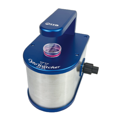
CytoWatcher II/CytoWatcher II FL is a digital microscope that can capture images of living cells and tissues for long periods of time without damaging them. The space-saving, power-saving design allows it to fit compactly inside a CO2 incubator, minimizing the impact on the environmental temperature. It supports not only brightfield photography but also fluorescent photography, and its highly reproducible color photography is also suitable for observing tissue sections and biological tissues.
Wound healing assay
A scratch (wound) was made in a confluent NIH3T3 cell with the tip, and the repair process was photographed at intervals of 10 minutes using CytoWatcher II. The dynamic cell migration can be observed.
Fluorescence imaging of GFP expressing cells
The image shows NIH3T3 cells 20 hours after transfection with an EGFP expression vector, photographed with a CytoWatcher II FL. Images were taken at intervals from 18 to 30 hours after gene transfection.
5-megapixel color CMOS sensor captures cell dynamics
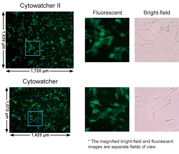
The CMOS image sensor used in CytoWatcher II has improved performance compared to conventional sensors, allowing it to capture clearer images than ever before. The sensor’s light-receiving section uses a back-illuminated and light-shielding structure, and the pixel size (area) is 1.6 times larger, which has improved sensitivity and saturation characteristics and reduced noise. In addition, the global shutter method (a sensor method that simultaneously captures the entire subject and then outputs it) is used, so even when shooting fast-moving subjects, the image is not distorted and high-precision images are obtained as they are.
The right figure shows images of GFP-expressing NIH3T3 cells taken with CytoWatcher FL and CytoWatcher II FL (left: 4x) and an enlarged image of the area enclosed in a square (center). The bright-field image is an image of the same cell in a different field of view enlarged in the same way as the fluorescent image (right).
The shooting area of CytoWatcher II FL has been expanded 1.6 times compared to the conventional CytoWatcher FL. The image resolution remains the same as the conventional model at 5 million pixels, but the larger pixel size allows for more sensitive shooting.
Photographed while installed in a CO2 incubator
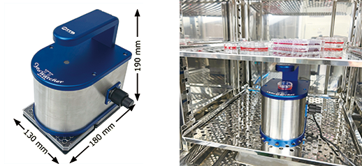
CytoWatcher II is a space-saving B6 size device with an installation area equivalent to two 96-well plates. It is short and very compact, so it does not take up much space even when installed in a CO2 incubator. It
is also moisture-proof, so it can be installed in a CO2 incubator with a high humidity environment of 95% or more and can be used for long-term imaging. The device only operates when imaging, turns on the light and the camera to take images. It does not become a source of unnecessary heat, and its impact on the environmental temperature is kept to a minimum.
Simply connect to the USB port on your PC to start recording.
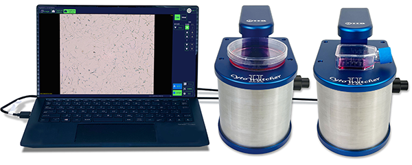
CytoWatcher II can be photographed simply by connecting it to a Windows PC with a USB 3.0 cable.
It is possible to connect two CytoWatcher IIs to one PC and photograph under the same photographing conditions and at the same time. This is a convenient function when performing experiments under multiple culture conditions or comparative experiments with controls.
Supports fluorescent imaging of living cells and tissue sections
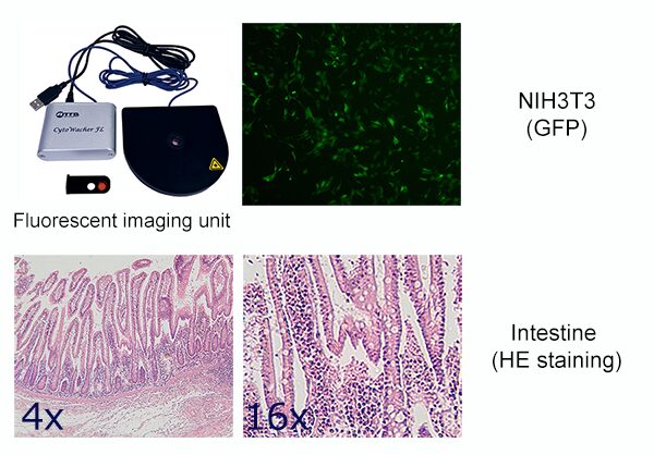
CytoWatcherⅡ FL is equipped with a blue LED light source (465 nm) for excitation, a short-pass filter (480 nm) for the excitation light source, and a band-pass filter (525/45 nm) for fluorescence imaging. It can capture green fluorescence such as GFP in living cells over time while they are still alive. It is also possible to capture bright-field and fluorescent images of the same cell in time-lapse mode.
In addition, the low-magnification lens provides a large field of view and higher resolution than a stereo microscope, allowing for precise image capture. The high-performance color CMOS sensor allows for highly reproducible color imaging similar to the color of actual tissues, such as animal tissue sections and plant tissues
Example of images taken by CytoWatcher
The data below are images taken with CytoWatcher (previous model). Similar images can be taken with CytoWatcher II.
Growth of NIH3T3 cells
Mouse fibroblast NIH3T3 cells on a 10 cm dish, Imaging for 72 h (30 minutes-intervals)
Differentiation into Adipocyte-like cells
Swiss 3T3 L-1 cells imaging for 7days (1 h-intervals) after replacing to the differentiation inducing medium (DMEM, 10 % FBS, 250 nM Dexamethazone, 0.5 mM Isobutylmethylxanthin, 10 ug/mL Insulin)
Wound healing assay
A scratch was made on the cell layer of mouse fibroblast NIH3T3 cells cultured to 100% confluence in a 35 mm dish, and images were taken at 30 minute intervals for 24 hours.
IVF bovine embryos
Time-lapse monitoring bovine embryos 6 hours after in vitro fertilization for 9 days (0.5 h-intervals) in an incubator
By courtesy of Kei Imai, Department of Sustainable Agriculture, Rakuno Gakuen University, Japan
Brochure
Specifications
| WSL-1850 CytoWatcher II | WSL-1850-B CytoWatcher II FL | |
|---|---|---|
| Camera | 5M pixels color CMOS camera | |
| Resolution | 2448 x 2048 pixels | |
| Magnification | Optical 4x (digital zoom: up to 16x) | |
| Field of view | 1.720 x 1.439 mm | |
| Focusing | Manual (coarse/fine) | |
| Light source | White LED (trans-illumination) | White LED (trans-illumination) 465 nm epi-blue LED (Side-illumination) |
| Optical filter | - | Excitation: 480 nm shortpass filter Emission: 525/45 nm bandpass filter |
| Moisture resistant | Usable in humidity 95% RH (usable in a CO₂ incubator) | |
| Control software | ImageSaverT for Windows (included as standard) Capture mode: Live / Time-lapse |
|
| File format | 8 bit TIFF, BMP, JPEG, Color / Monochrome Movie file: avi, maximum 60 fps |
|
| PC connection | USB 3.0 x 1 | USB 3.0 x 1, USB 2.0 (or 3.0) x 1 |
| OS | Windows 10/11 (64/32 bit) | |
| Dimension | 130 (W) x 180 (D) x 190 (H) mm | 130 (W) x 180 (D) x 190 (H) mm |
| Weight | 2.5 kg | 2.9 kg |
| Power | USB (bus powered) | |
Ordering Information
| Code No. | Description | Unit |
|---|---|---|
| 3601850 | WSL-1850 CytoWatcher II | 1 set |
| 3601853 | WSL-1850-B CytoWatcher II FL | 1 set |
| 3601856 | Fluorescent imaging unit B for WSL-1850 | 1 pk |
| 3601809 | USB 3.0 extension cable | 1 pk |
